
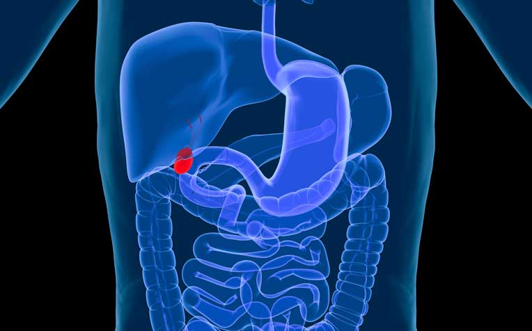
Biliary Tract Disease
Introduction to Gallbladder & Biliary Tract Diseases
|
Biliary disease is diseases affecting the bile ducts, gallbladder and other structures involved in the production and transportation of bile. Bile is a fluid produced by the liver that aids digestion. Bile drains from the liver through bile ducts to the first part of the small intestine, or duodenum, and eventually back to the bile ducts and liver. If any duct in this complex system becomes diseased or blocked, a number of serious diseases—discussed below—may result (The George Washington University Hospital Opens in new window). |
- Gallstones
The most frequent cause of gallbladder disease is gallstones (95%). It is more common in women than men, and the incidence increases with age. Recurring episodes of symptoms are characteristic. Gallbladder disease is diagnosed by a combination of clinical features, laboratory investigations and organ imaging.
Patients present with biliary pain caused by obstruction of biliary flow, leading to dilation of the biliary system. Cholecystitis and ascending cholangitis develop when secondary infection occurs. Calculous disease is the most frequent cause of pancreatitis. Acalculous cholecystitis occurs in the absence of gallstones and may complicate major illness .
1.1 Clinical Features
1.1.1 History
Patients usually present with abdominal pain which may be midline and visceral or somatic and right upper quadrant. Visceral pain may be referred around the right costal margin or to the right shoulder area. Despite the use of the term biliary colic, the pain is usually constant and may be severe. Nausea and vomiting are often present. Complaints of fevers and chills may be indicative of either cholecystitis or ascending cholangitis. Rigors are suggestive of cholangitis.
1.1.2 Examination
Right upper quadrant tenderness is the most common examination finding. Fever and tachycardia are usually present in acute cholecystitis, although at presentation they may be absent in 59–90% of cases. Local peritonism and Murphy’s sign also suggest acute cholecystitis. Jaundice is usually absent in biliary colic and acute cholcystitis. The presence of pain, jaundice, high fever and shaking chills (Charcot’s triad) is indicative of ascending cholangitis.
1.2 Differential Diagnosis
The differential diagnosis of right upper quadrant pain includes:
- Peptic ulcer disease, including perforation.
- Acute pancreatitis.
- Coronary ischaemia, especially involving the inferior myocardial surface.
- Appendicitis, especially retrocaecal or in pregnancy.
- Renal disease, including renal colic and pyelonephritis.
- Colonic conditions.
- Hepatic pathology, especially hepatitis.
- Right lower lobe pneumonia.
1.3 Investigations
Investigation of biliary pain are aimed at confirming the diagnosis, establishing the presence of gallstones and the detection of complications.
1.3.1 Imaging
Ultrasound is the investigation of choice to confirm the diagnosis and measure the thickness of the gallbladder wall and the diameter of the common bile duct (CBD). It can also detect the presence of calculi in the CBD and the presence of any local fluid collection. It has high sensitivity and specificity, is non-invasive, and requires little preparation of the patient. It does, however, require experience in technique and interpretation.
In the majority of cases plain radiographs are not helpful in the diagnosis of gallbladder disease, but on occasion they may be useful to rule out other potential diagnoses. Rare X-ray findings include radio-opaque calculi (only 10–15% of biliary calculi are radio-opaque), the presence of gas in the biliary tree indicating a biliary-gastrointestinal fistula, gas or an air-fluid level in emphysematous cholecystitis, or a localized ileus in the right upper quadrant.
1.3.2 Blood Tests
Blood tests are relatively non-specific. Bilirubin and alkaline phosphatase levels are mildly elevated in uncomplicated biliary colic and cholecystitis. Amylase and lipase are elevated if pancreatitis is also present. Full blood examination shows a leukocytosis and left shift in the majority of cases of cholecystitis and cholangitis; however, 32–40% do not have a leukocytosis.
1.4 Complications
Complications of biliary disease include:
- Cholecystitis
- Obstructive jaundice
- Ascending cholangitis and Gram-negative septicaemia
- Gallstone ileus
- Perforation: the elderly and diabetics are at particular risk of rapid necrosis and perforation
- Pancreatitis.
1.5 Management
The management of biliary pain depends on the presence or absence of complications. In the emergency department (ED) phase patients should receive analgesia in the form of titrated intravenous opioids, and intravenous fluids.
In selected patients, parenteral non-steroidal anti-inflammatory drugs (NSAIDs) may be effective. There is some evidence that a short course of NSAIDs may prevent progression to cholecystitis in some patients with biliary colic. In the absence of cholecystitis or complications such as biliary obstruction, ascending cholangitis or pancreatitis patients may be discharged for outpatient surgical follow-up if the pain settles.
Antibiotics are indicated for the treatment of cholecystitis or ascending cholangitis. The appropriate antibiotics for cholecystitis in which Gram-negative organisms are most frequently implicated are ampicillin and gentamicin, or cefotaxime if the patient is penicillin allergic. Ascending cholangitis should be treated with cefotaxime or ceftriaxone.
Endoscopic retrograde cholangiopancreatography (ERCP) is indicated for the treatment of biliary obstruction. Surgical removal of gallstones is indicated for all patients who are fit for the procedure. The timing of surgery is a matter of surgeon preference and theatre availability.
1.6 Disposition
Many patients with biliary colic can be discharged. Most patients with complications such as acute cholecystitis, ascending cholangitis or pancreatitis require hospital admission. Admission may also be indicated in some cases because of recurrent severe pain.
- Cholelithiasis
2.1 Epidemiology
The most common abdominal pathology leading to hospital admission in developed countries is cholelithiasis. Gallstones are present in 10–20% of the adult population in developed countries, but more than 80% are ‘silent’. In developed countries, the majority of gallstones are formed predominantly from cholesterol (up to 80%). Increased age is associated with lithogenic bile and an increased rate of gallstones. In young adults, more females are affected than males, but the disparity narrows with age.
The lifetime risk of cholesterol gallstones is 35% in women, compared to 20% in men. This is likely to be due to endogenous sex hormones that enhance cholesterol secretion and increase bile cholesterol saturation. In addition, progesterone may contribute by relaxing smooth muscle and thereby impairing gallbladder emptying.
Other than older age and female gender, predisposing factors include obesity, a high-calorie diet, total parenteral nutrition, weight loss (especially if rapid), drugs (including clofibrate, oral contraceptives and other exogenous oestrogens and ceftriaxone), genetic predisposition, diseases of the terminal ileum and abnormal lipid profile.
Pregnancy is also a predisposing condition. Gallstone precipitation is common, especially in late pregnancy, but most remain asymptomatic, at least until delivery. Symptomatic cholelithiasis can complicate the puerperium and each first postnatal year. Forceful gallbladder contraction postpartum increases the potential for cystic or common bile duct obstruction.
2.2 Clinical Features and Investigation
Many gallstones are present for decades before symptoms develop and 70–80% remain asymptomatic throughout life. Asymptomatic patients convert to symptomatic at a rate of 1–4% per year (the risk decreases with time).
The most common presentations are biliary colic, cholecystitis, obstructive pancreatitis (5% of all patients) and ascending cholangitis. Less common presentations are empyema, perforation, fistula formation, gallstone ileus, hydrops or mucocoele of the gallbladder and carcinoma of the gallbladder.
The investigation of choice is ultrasound, which has a sensitivity of 84–97% and a specificity of 95–100%.
2.3 Treatment
Cholecystectomy is the definitive treatment of choice for symptomatic cholelithiasis. It provides symptomatic relief in up to 99% of patients. Laparoscopic cholecystectomy is the technique of choice.
Dissolution methods and lithotripsy are of limited utility owing to restricted indications for their use and gallstone recurrence at 5 years in approximately 50% of cases. Prophylactic cholecystectomy is not recommended in asymptomatic patients as the risks of the procedure outweigh the potential benefits.
- Acute Cholecystitis
3.1 Epidemiology
Distribution parallels that of cholelithiasis. Acute cholecystitis develops in 1–3% of patients with symptomatic stones.
3.2 Pathology
Cholelithiasis is present in most acute cases, a single large calculus being the most common finding. A small group of patients develop biliary sludge, which is a mixture of particulate matter and bile. More than 90% of cases result from cystic duct obstruction.
Bacteria are present in approximately 20–50% of case, but bacterial infection is not thought to cause acute cholecystitis; rather, it results from chemical irritation an inflammation of the obstructed gallbladder due to obstruction of the cystic or common bile duct. Secondary bacterial infection is usually caused by aerobic bowel flora (such as Escherichia coli klebsiella species and, less commonly, Enterococcus faecalis). Anaerobes are found infrequently, usually in the presence of obstruction.
3.3 Clinical Features
Right upper quadrant pain and fever are the most common features. Usually patients have experienced previous episodes of biliary pain. Nausea and vomiting are commonly present. A distended, tender gallbladder is not usually evident: the right upper quadrant mass palpated in approximately 20% of patients represents omentum overlying the inflamed gallbladder. Only approximately 20% of patients are jaundiced. The presence of hyperbilirubinaemia suggests common bile duct obstruction. Neutrophilia may be present.
3.4 Imaging
3.4.1 Ultrasound
Findings on ultrasound are often diagnositic, showing cholelithiasis, an increase in transverse gallbladder diameter >4–5 cm in up to 87% of cases, gallbladder wall thickening >5 mm and pericholecystic fluid. A positive Murphy’s sign on ultrasound is a sensitive indicator of cholecystitis.
3.4.2 Plain abdominal X-rays
Abdominal X-rays are rarely helpful, but in 10% of cases may show radio-opaque gallstones. In emphysematous cholecystitis, gas may be seen within the gallbladder wall.
3.5 Complications
Complications include bacterial superinfection leading to ascending cholangitis or sepsis, gallbladder perforation leading to local abscess formation or diffuse peritonitis, biliary enteric (cholecystenteric) fistula, with a risk of gallstone-induced intestinal obstruction (gallstone ileus), and deterioration in pre-existing medical illness.
3.6 Treatment
Treatment is with antibiotics, hospital administration and cholecystectomy. (Amoxy)ampicillin 1 g i.v. 6-hourly, plus gentamicin at 4–6 mg/kg i.v. daily is recommended (the latter should be adjusted for decreased renal function). If these are contraindicated the alternatives are ceftriaxone 1 g i.v. daily or cefotaxime 1 g i.v. 8-hourly.
It is important to note that cephalosporins are not active against enterococci. In the presence of biliary obstruction, metronidazole should be added to treat anaerobes.
Most patients will respond to conservative management, with the gallstone disimpacting and falling back into the gallbladder, thereby allowing the cystic duct to drain. If the gallstone does not disimpact, gangrenous cholecystitis (2–30% of cases), empyema of the gallbladder or gallbladder perforation (10% of cases) may occur.
Cholecystectomy is required to prevent recurrence or other complications. Approximately 20% of patients require emergency surgery, usually via a laparoscopic approach. If performed within 72–96 hours of the onset of symptoms, the surgery is easier and complication rates are lower. The timing of cholecystectomy for the remaining 80 will be determined by the treating surgical unit.
- Acalculous Cholecystitis
Acute acalculous cholecystitis is acute inflammation of the gallbladder in the absence of gallstones, generally in the severely ill patient, and accounts for 10% of cases of acute cholecystitis. Predisposing factors include postoperative state after major, non-biliary surgery, severe trauma or burns, multi-system organ failure, sepsis, prolonged intravenous hyperalimentation, and the postpartum state.
It is thought to be ischaemic in pathologenesis, with more than 70% of patients having underlying atherosclerotic disease. Contributing factors include dehydration, multiple blood transfusions, gallbladder stasis, accumulation of biliary sludge, viscous bile and gallbladder mucus, and bacterial contamination. Compared with acute calculous cholecystitis, there is a much higher incidence of empyema, gangrene and perforation of the gallbladder, and consequently an increased mortality rate (up to 50%).
- Choledocholithiasis
5.1 Features
Gallstones are present within the biliary tree, almost all derived from the gallbladder. Approximately 10% of patients with cholelithiasis have choledocholithiasis, which may be asymptomatic, intermittently or permanently obstructive. Choledocholithiasis is the second most common cause of CBD obstruction after neoplasms.
Symptomatic cases present due to obstruction (resulting in jaundice), pancreatitis, cholangitis, hepatic abscess, secondary biliary cirrhosis or acute acalculous cholecystitis.
5.2 Imaging and Treatment
Ultrasound is less reliable in choledocholithiasis. CBD measurement may yield false positive or false negative results, but is more accurate in jaundiced patients, approaching 80% accuracy. In addition, the precise level and cause of obstruction is sometimes difficult to identify, especially if it lies near the pancreatic head.
ERCP is more accurate and often therapeutic. Interval cholecystectomy to prevent recurrence is recommended. The management of asymptomatic duct calculi is controversial. Options inclue laparoscopic cholecystectomy with endoscopic sphincterotomy and stone extraction, or laparoscopic exploration of the common bile duct.
- Cholangitis
6.1 Aetiology
Cholangitis is a purulent bacterial infection of the biliary tree, including the intrahepatic ducts related to obstruction to bile flow (e.g. choledocholithiasis, stents, tumors, acute pancreatitis and strictures). Parasitic infections are a rare cause in developing countries but are common in developing countries. Bacteria are usually Gram-negatives such as E. coli, Klebsiella, Clostridium spp, Bacteroides and Enterobacter or group D streptococci. They are thought to enter the biliary tree via the sphincter of Oddi.
Charcot’s biliary triad (fluctuating jaundice, recurrent right upper quadrant abdominal pain and intermittent high fever with rigors) is present in 70% of patients.
6.2 Treatment
The principles of treatment are broad-spectrum antibiotics and prompt drainage of the obstruction. For the latter, the method will depend on the underlying cause, surgical preference and availability, and the state of the patient.
The recommended antibiotics are (amoxy)ampicillin 2 g i.v. 6-hourly, plus gentamicin 4–6 mg/kg i.v. daily. Metronidazole 500 mg i.v. 12-hourly should be added in patients with a history of previous biliary tract surgery or known biliary obstruction. If these are contraindicated the alternatives are ceftriaxone 1 g i.v. daily or cefotaxime 1 g i.v. 8-hourly. Delay in management may lead to septicaemia and hepatic abscess formation, which are associated with a high mortality.
See also:
- Adapted from: Textbook of Adult Emergency Medicine E-Book. Authored By Peter Cameron, George Jelinek, Anne-Maree Kelly, Lindsay Murray, Anthony F. T. Brown. Further Readings as cited include:
- Beckingham IJ. ABC of diseases of liver, pancreas, and biliary system: gallstone disease. British Medical Journal 2001; 322: 91–94.
- Beckingham IJ, Ryder SD. ABC of diseases of liver, pancreas, and biliary system: investigation of liver and biliary disease. British Medical Journal 2001; 322: 33–36.
- Cotran RS, Kumar V, Collins T. Robbins pathologic basis of disease, 6th edn. Philadelphia: WB Saunders, 1999.
- Epstein FB. Acute abdominal pain in pregnancy. Emergency Medical Clinics of North America 1994; 12: 151–165.
- Feldman M, et al. Sleisenger and Fordtran’s gastrointestinal and liver disease, 6th edn. Philadelphia: WB Saunders, 1998.
- Gruber PJ, Silverman RA, Gottesfeld S, et al. Presence of fever and leukocytosis in acute cholecystitis. Annals of Emergency Medicine 1996; 28: 273–277.
- Hudson PA, Promes SB. Abdominal ultrasonography. Emergency Medical Clinics of North America 1997; 15: 825–848.
- Indar AA, Beckingham IJ. Acute cholecystitis British Medical Journal 2002; 325: 639–643.
- Johnson CD. ABC of the upper gastrointestinal tract: upper abdominal pain — gall bladder. British Medical Journal 2001; 323: 1170 –1173.
- Kumar A, Deed JS, Bhasin B, et al. Comparison of the effect of diclofenac with hyposcine-N-butylbromide in the symptomatic treatment of acute biliary colic. American New Zealand Journals of Surgery 2004; 74: 573–576.
- Moscati RM. Cholelithiasis, cholecystitis and pancreatitis. Emergency Medical Clinics of North America 1996; 14: 719–737.
- Singer AJ, McCracken G, Henry MC. Correlation among clinical, laboratory and hepatobiliary scanning findings with acute cholecystitis. Annals of Emergency Medicine 1996; 28: 267–272.
- Therapeutic Guidelines (Australia): Antibiotic. Version 11. Therapeutic Guidelines Limited, 2007. Accessed at http://etg.hcn.net.au/ December 2007.