
Electroencephalography
 Graphics courtesy of HealthlineOpens in new window
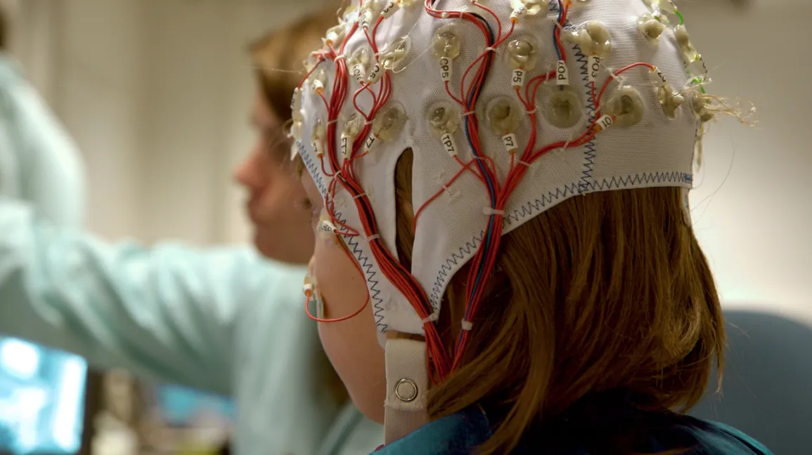
Electroencephalograpy (or EEG) is a measure of brain activity. The procedure involves attaching electrodes to an individual’s scalp. The electrodes measure electrical activity of brain cells close to the scalp and provide millisecond-by-millisecond readings. |
An EEG can be used by presenting a visual, auditory, olfactory, or other stimulus to an individual and recording the EEG changes that occur after the stimulus was presented. The readings produce specific information about time of experience but not always about specific location of the brain activity, although general location is indicated. Compared to most measures of brain activity, EEGs are inexpensive to use.
An EEG can be helpful in providing information about emotionOpens in new window; however, brain activity is recorded only in areas close to the sacp, whereas may brain areas relevant to emotion are located in the interior.
Despite the shortcomings associated with using EEGs, some important information about emotion has been discovered or confirmed through their use. For instance, a volume of EEG evidence shows that the right hemisphere of the brain tends to be associated with negative affect (immediate, physiological response and evaluation of a stimulus as good or bad) and the left with positive affect (e.g., Davidson, 2003).
As another example, using both EEG and functional magnetic resonance imaging (fMRI), Seitz et al. (2008) found that the subjective experience of empathy, as a response to observing another person’s facial expression of empathy, involves the activity of the dorsal medial frontal cortex of the brain.
- Davidson, R. J. (2003). Affective neuroscience and psychophysiology: Toward a synthesis. Psychophysiology: 40, 655 – 665.
- Seitz, R.J., Schaefer, R., Scherfeld, D., Freiderichs, S., Popp, K., Wittsack, H.-J., et al. (2008). Valuating other people’s emotional face expression: A combined functional magnetic resonance imaging and electroencephalography study. Neuroscience, 152, 713 – 722.
