
Ephelis (Ephelides "plural")
Clinical Definition
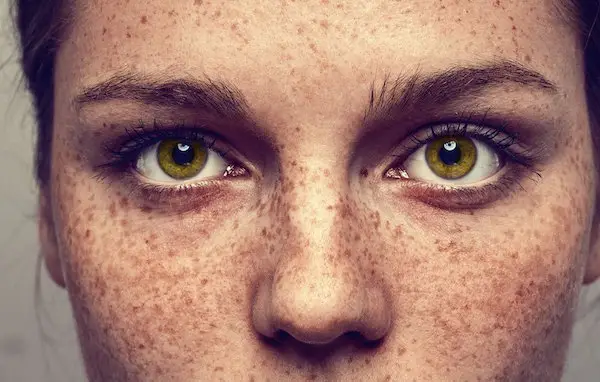 Figure X-1 | Photo courtesy of Salon Compass Opens in new window
Ephelides are benign clusters of small, well-circumscribed, focal, pigmented lesions found only on sun-exposed skin in which despite their increased and persistent pigment synthesis, the constituent melanocytes Opens in new window do not appear to have acquired the neoplastic characteristics of nevus cells Opens in new window.
Ephelides (also known as freckles) typically manifest in childhood in fair-skinned (phototypes I and II) individuals, especially those with red or blonde hair color.
Ephelides tend to be passed down from generation to generation in an autosomal dominant inheritance pattern. However, the tendency to develop ephelides decreases with advancing age.
Clinical Features
Ephelides occur at a very young age and are accentuated in sun-exposed regions of the body, particularly the face, the dorsal aspects of the arms, and the upper part of the chest and the back.
Exposure to the sun or other ultraviolet source causes the ephelides to become darker and clinically more noticeable. They do not occur within the oral mucosa.
They are usually uniform in coloration but can have many different sizes and shapes. Some are round or oval; others are angulated or have a bizarre shape; and are usually 1–3 mm in diameter, but some may be several millimeters in diameter.
Dependent on the intensity of sun exposure, their color is usually a uniform light to dark brown but almost never become as black as lentigines Opens in new window or junctional nevi Opens in new window; they may increase in number and distribution and show a tendency to confluency.
Patients with multiple ephelides may have a higher risk for skin cancer, because their presence may be an indication of increased exposure to ultraviolet radiation.
As mentioned earlier, ephelides become less prominent with advancing age, but even when clinically inapparent in older persons with ordinary light, they remain visible upon Wood’s lamp examination.
Pathogenesis
Ephelides are thought to be genetically inherited, most likely in a dominant pattern. They become more pronounced with sun exposure and fade during times with less exposure to ultraviolet radiation.
The increase in pigment is caused by an increase in the production of melanin and an increase in the transfer of melanosomes from melanocytes to keratinocytes. There is no increase in the number of melanocytes Opens in new window in ephelides.
Recently a significant association has been shown between melanocortin-1-receptor (MCIR) gene variants and the propensity to develop ephelides.
The carriers Opens in new window of one or two MCIR gene variants (there are currently some 27 MCIR gene variants) demonstrated a 3- and 11-fold risk for developing freckles, respectively (both p<0.0001).
The latter relationships are independent of skin phototype and hair color and are comparable in individuals with and without a history of skin cancer.
Differential Diagnosis
Histologically, ephelis (singular) cannot be distinguished from café-au-lait macule Opens in new window, melanotic macule of Albright, and melasma Opens in new window.
Discrimination of ephelis (or freckle) from the latter entities must be based solely on clinical findings.
The solar and simple lentigines also may enter into the differential diagnosis of ephelis.
The simple lentigo Opens in new window differs from the freckle by showing elongated epidermal rete ridges and melanocytic hyperplasia.
The solar lentigo usually has prominently club-shaped rete, or a flattened or effaced epidermal silhouette; increased frequency of basilar melanocytes may or may not be present.
Therapeutic
No therapy is required other than to recommend sun protection, sunscreen use, and routine skin examinations in the future.
