
Idiopathic Guttate Hypomelanosis
Medical Definition
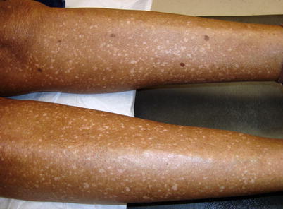
Idiopathic guttate hypomelanosis (IGH) is a common benign acquired leukoderma that is characterized by discrete, well-define, round white macules on the sun-exposed areas of the legs and forearms. The patches are usually asymptomatic and can resemble vitiligo Opens in new window.
Epidemiology
The incidence of IGH which increases with age, is seen in up to 80% of patients over the age of 70 years. Although IGH is more common patients with advanced age, it has also been observed in young adults. IGH occurs in all races and skin types, but it is more striking in darkly pigmented skin.
In Caucasians, IGH may favor those with brown eyes and brown hair. Female incidence appears to be higher, but this could be due to the fact that women are more likely to seek medical attention for aesthetic reasons.
Etiology
The etiology and pathogenesis of IGH are unknown. IGH has been hypothesized to be UV-induced, as it most commonly affects sun-exposed sites, such as the extremities, neck, and face.
Yet another hypothesis for the etiology of IGH is normal aging or photoaging. Other suggested contributing factors include genetics, trauma, and autoimmunity.
Clinical Features
Clinically, IGH consists of small hypopigmented and depigmented macules which are sometimes porcelain white, with discrete circumscribed borders (as depicted in the image above).
The macules of IGH measure about 0.5–6 mm in diameter and often delineated by the skin furrows; occasionally, macules are up to 2.5 cm in size. They are often scattered and usually observed on the exposed areas of the lower extremities. In most patients, there are multiple macules on the extensor forearms and shins; the remainder of the extremities can be affected, but rarely the face.
Natural History and Prognosis
Once present, the macules of IGH may grow slightly but do not change much in size. The lesions do not coalesce; their surface is smooth but not atrophic.
Spontaneous repigmentation has not been reported. Hairs within the macules usually retain their pigment. Although benign and asymptomatic, affected individuals may seek medical attention for aesthetic reasons.
Histopathological Features
The most consistent histologic features of IGH are a flattening of the dermal-epidermal junction, moderate to marked reduction or focal absence of melanin granules in the basal and suprabasal layers, and a basket-weave hyperkeratosis.
There is a moderate to relatively marked reduction in the number of DOPA-positive epidermal melanocytes (10-50% compared with normal skin), but these cells are never totally absent. At the ultrastructural level, some melanocytes have normal melanogenic activity, while others lack mature melanosomes.
Diagnosis and Differential Diagnosis
IGH can mimic several skin disorders of pigmentation. The differential diagnosis includes post-inflammatory hypopigmentation Opens in new window, vitiligo Opens in new window, lichen sclerosis, pityriasis lichenoides chronic, pityriasis alba Opens in new window, atrophie blanche, leprosy Opens in new window, leukoderma following PUVA therapy, and confetti-like lesions of tuberous sclerosis.
In the neoplastic category, the differential diagnosis includes hypopigmented mycosis fungoides, disseminated hypopigmented keratoses following PUVA therapy, and achromic verruca plana with the latter two entities forming papules to help distinguish them from IGH.
Therapeutics
IGH does not require treatment. Patients who seek the attention of a dermatologist may choose to explore therapy for aesthetic or cosmetic reasons after being reassured of the benign nature of this condition.
Multiple therapies are available all with variable success rates of achieving the patient’s desired repigmentation. These include cryotherapy, superficial dermabrasion, topical retinoids, fractional carbon dioxide lasers, intralesional corticosteroids, and topical tacrolimus and pimecrolimus.
See also:
- Kim SK, Kim EH, Kang HY, Lee ES, Sohn S, Kim YC. Comprehensive understanding of idiopathic guattate hypomelanosis: clinical and histopathological correlation. Int J Dermatol. 2010;49(2):162-6.
- Shin J, Kim M, Park SH, Oh SH. The effect of fractional carbon dioxide lasers on idiopathic guttate hypomelanosis: a preliminary study. J Eur Acad Dermatol Venereol. 2013;27(2):e243-6.
- Rerknimitr P, Disphanurat W, Achariyakul M. Topical tacrolimus significantly promotes repigmentation in idiopathic guttate hypomelanosis: a double-blind, randomized, placebo-controlled study. J Eur Acad Dermatol Venereol. 2013;27(4):460-4.
- Falabella R, Escobar C, Giraldo N, et al. On the pathogenesis of idiopathic guttate hypomelanosis, J Am Acad Dermatol. 1987;16(1 Pt 1):35-44.
- Ortonne J-P. Vitiligo and other disorders of hypopigmentation. In:Bologna JL, Jorizzo JL, Rapini RP, editors. Dermatology, vol. 1. 2nd ed. St. Louis: Mosby; 2008. P. 935.
- Hexsel DM. Treatment of idiopathic guttate hypomelanosis by localized superficial dermabrasion. Dermatol Surg. 1999;25(11):917-8.
- Friedland R, David M, Feinmesser M, Fenig-Nakar S, Hodak E. Idiopathic guttate hypomelanosis-like lesions in patients with mycosis fungoides: a new adverse effect of phototherapy. J Eur Acad Dermatol Vereol. 2010;24(9):1026-30.
- Calonje E. Brenn T, Lazar A, McKee PH. Disorders of pigmentation. In: Calonje E, Brenin T, Lazar A, McKee PH, editors. McKee’s pathology of the skin with clinical correlations, vol. 2. Printed in China. Edinburgh: Elsevier/Saunders, 2012. http://www.ncbi.nlm.nih.gov/nlmcatalog/1015484719.
- Ploysangam T. Dee-Ananlap S, Suvanprakorn P. Treatment of idiopathic guttate hypomelanosis with liquid nitrogen: light and electron microscopic studies. J AM Acad Dermatol. 1990;23(4 Pt 1):681-4.
