
Junctional Nevus
Clinical Definition and Features
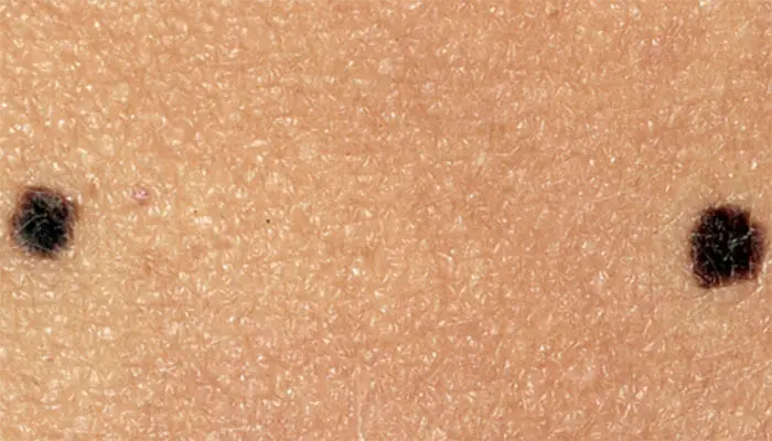 Figure X-1: Two junctional nevi that appear as uniformly brown small macules, round in shape with smooth regular borders | Photo courtesy of WebMD Opens in new window
Junctional nevus is defined as a nevus in which the melanocytic proliferation is restricted to the basal portion of the epidermis (‘junctional’ area). Microscopically, it is characterized by the presence of melanocytic nests (‘theques’) on the epidermal side of the dermoepidermal junction.
Nevi of the palms and soles are nearly always of the junctional type, with most of the intraepidermal melanocytes concentrated in the skin furrows.
Clinical Features
From a clinical point of view junctional nevus is a small flat, or slightly elevated, nonhairy, uniformly pigmented, dark brown to almost black lesion with a smooth surface (Figure X-1).
Its lateral diameter does not exceed 6 millimeters. It is typical of young patients and can occur on any site of the body. Occasionally, junctional nevi are very numerous, usually they are 20–30.
As junctional nevi enlarge, there is an increasing tendency for them to acquire an irregular border, variability in color and other features that result in a biopsy.
The dividing line between the “ordinary” junctional nevus and a junctional “dysplastic” nevus may rest on the clinical features in some cases, on the histopathologic ones in others, and may be largely arbitrary.
Histological Features
Histologically, this nevus has two major aspects:
- The proliferation of melanocytes along the dermoepidermal junction
- The collection of the melanocytes in roundish nests.
These two aspects are briefly discussed below.
- Proliferation of junctional melanocytes
An increased number of melanocytes in single unit is present at the junction. Cells are morphologically typical and colonize the basal layer of the rete ridges, which become elongated and thickened.
Usually, the suprapapillary plate is less involved by the melanocytic proliferation (or is altogether undisturbed by it).
The proliferating melanocytes are usually roundish and epithelioid with no dendrites; rarely they retain their dendrites and have an elongated hyperchomatic nucleus.
- Collection of the melanocytes in roundish nests (aggregation in nests)
Melanocytes aggregate in the theques that are round clusters mostly found at the bases of elongated rete ridges.
Nests are uniform in size and shape and evenly distributed throughout the lesion. Characteristically, two nests delimit the lateral borders of the lesion, which “starts with a nest and ends with a nest”.
- Melanocytes in the nests usually lack dendrites and have small round nuclei with dispersed chromatin
- A punctuate nucleolus is usually distinguishable
- The nuclear membrane is thin and homogenous without clumped chromatin
- Cytoplasm is scarce and contains coarse melanin granules
- Cells are monomorphic and uniform in size and shape
- When the pigment is abundant inside the nests, scattered melanophages are also presnt.
In the papillary dermis a band of melanophages is present in heavily pigmented lesions (as a rule, however, junctional nevus is less pigmented than lentigo simplex).
The (supposed) natural history of a junctional nevus consists in a progressive “maturation” first into a compound Opens in new window and then into an intradermal nevus Opens in new window. This is not obligatory: a junctional nevus can remain indefinitely in its original form.
Variants
- Hypermelanotic nevus
This lesion (Cohen) is darkly pigmented and frequently excised for its worrisome clinical appearance. The salient characteristic is the abundance of melanin.
Melanin granules are present in the basal layer where they blur the nuclear and cytoplasm details of the keratinocytes.
Melanin is present in corneocytes too, where it aggregates in small round spheres, which simulate dark nuclei creating “pseudo-parakeratosis”.
The melanocytes have elongated and pigmented dendrites which insinuate themselves among the keratinocytes.
Dendrites are also retained in the nests where they form a thick blackish net enveloping small, irregularly shaped, nuclei.
Cells inside the nests are more pigmented than those disposed in single units along the basal layer.
Melanocytes are mostly single; only few nests are present. This variant of junctional nevus is similar to the so called hypermelanotic lentigo.
- Junctional nevus with epithelioid cells
This is a variant of junctional nevus with large epithelioid melanocytes. Patients are usually adolescent, rarely adults.
Melanin can be copious with many melanophages scattered in the papillary dermis. Large multinucleated melanocytes are also a finding. This variant can be confused with a form of melanoma if more than an occasional atypical nucleus is found.
