
Correlational Studies
.webp) File photo. The Developmental Stages of a Child (Online Psychology Degree Guide, 2021)
File photo. The Developmental Stages of a Child (Online Psychology Degree Guide, 2021) |
Let us begin with a definition: a correlation coefficient is a statistic between +1 and –1 which indicates the extent to which two variables tend to be related or to vary together. A value close to +1 is a high positive correlation which tells us that the two variables are closely related.
There are many instances of naturally occurring positive correlations: between height and weight (taller people tend to weigh more); between maths and English (students tend to be good, bad or indifferent at both!) There are innumerable instances of correlations that are close to zero (indicating no relationship): height is not correlated with academic performance; IQ is not correlated with sports achievement.
A correlation coefficient close to –1 is a high negative correlation which tells us that two variables are inversely related. There are fewer instances of negative correlations — perhaps amount of time spent watching television and school grades!
There are primarily two types of correlational studies that are of interest to the developmental psychologists, concurrent and predictive.
- Concurrent studies
A concurrent correlational study is where we are interested in the relationship between variables that are measured at the same time.
An example of such a study would be to find out how similar the IQs of identical twins are. In this study we would give intelligence tests to pairs of identical twins and, if the correlation is high (which it almost certainly would be) this would tell us that if one twin had a high IQ the other one would also be bright; if one had a low IQ we could predict a low IQ for the other twin.
- Predictive studies
A predictive correlational study is one where we are interested in finding whether individuals retain their relative standing, or rank order, relative to others, over time.
For example, does the bright child at age 5 turn out to be the gifted student at age 20?; does the outgoing child become an extraverted teenager?
Here is one example of a predictive correlational study, asking the question ‘can we predict IQ in 3-year-olds from problem-solving in infancy?’
This is a study carried out by Peter Willatts, a developmental psychologist at Dundee University, Scotland, UK. It begins with 9-month-old infants who were tested on what is called a means-end problem-solving task. Each infant was shown an attractive toy which was placed out of reach on a cloth, and their job was then to grasp the cloth, pull it towards them (the mean) in order to take the toy (the end). This doesn’t sound too difficult, but babies only begin to string behaviours together to solve means-end tasks around 7 or 8 months – at 9 months many can do it expertly (see Figure X-1), but others are lagging behind.
Willatts then gave the same infants the British Picture Vocabulary Test (BPVT) when they were 3 years 3 months old (the BPVT is the British version of the well-known American test the Peabody Picture Vocabulary Test, PPVT, which is a test of intelligence).
What Willatts found was that those 9-month-old infants who were best at the means-ends task tended to become the 3-year-olds with the higher IQs.
The correlation was 0.64, and the relationship between the infants’ scores at 9 months and their scores as children is shown in Figure X-1. This figure is called a scattergram and is a graphical way of showing a correlation.
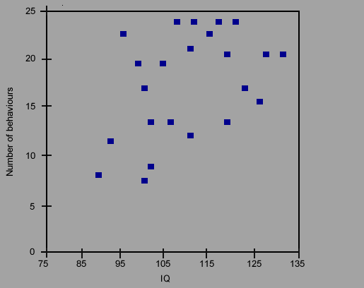 Figure X-1 Scattergram to show the relationship between the number of successful reaching behaviours at 9 months (vertical axis) and 3-year IQ (horizontal axis). The most successful infants turned out to be those with the higher IQs.
Source: Peter Willatts (1997). |
Correlational studies are thus important in telling us what sorts of abilities or psychological characteristics tend to go together (concurrent studies) and what abilities and characteristics predict later occurring behaviours (predictive studies).
- Neurodevelopmental studies
A particularly challenging task for developmental psychologists is to understand brain development and its relation to developments in perceptual, cognitive, social and motor skillsOpens in new window. This challenge is particularly acute for developmentalists because our subjects of interest—infants and children—can be difficult to test due to a general lack of cooperation or inability to cope with the methods, and also because the brain develops at a rapid pace early in life, making brain-behaviour links difficult to assess. Nevertheless, progress is being made with the judicious use of selective methods.
- Marker tasks
A marker task refers to a method designed to elicit a behaviour with a known neural basis. Often the neural basis is discovered through experiments with animals, for which experiments on brain function present fewer ethical hurdles than experiments with humans. For example, much is known about the neural basis of visual function from experiments with monkeys.
Visual attention in monkeys has been a subject of much investigation, and the neural underpinnings of different kinds of eye movements is fairly well known. It is thought that the visual system of rhesus macaques, a species of Old World monkeys, has a great deal of similarity to the human visual system, and researchers interested in the development of visual attention in humans have looked to the literature on monkeys for clues.
Marker tasks have contributed much to this goal (Johnson, 1990). One prominent example comes from infants’ ability to track moving objects using ‘smooth pursuit’ eye movements in which the point of gaze stays more or less locked on target as an object moves back and forth. Before 1 – 2 months of age, infants’ tracking is jerky and frequently falls behind the object, necessitating numerous attempts to catch up to it.
Johnson (1990) proposed that a specific area of the visual system known as the medial temporal (MT) area in the monkey has an analogue in the human, and development of this area and its connections with other parts of the visual system is responsible for the onset of smooth pursuit in humans. This is because damage to monkey MT causes deficits in motion tracking.
- Imaging methods
Recording brain activity in any animal poses a host of technical challenges, and no animal presents more of a challenge in this respect than a human child! Nevertheless, there are several methods available.
There are two kinds of imaging used commonly with infants and children: those that record brain activity from the scalp, and those that record activity inside the head. Scalp recordings are done with electrodes that measure electrical activity produced by neurons, yielding an electroencephalogram, or EEG.
The EEG is often measured when it is time-locked to a stimulus event, producing an event-related potential, or ERP. ERPs to different events can be compared to investigate developmental changes or individual differences in response.
The EEG and ERP is highly sensitive to the timing of the brain’s response to events, but it can be difficult to tap into specific brain regions with this method, because all activity is recorded at the surface. Accessing deep structures, such as those areas involved in memory or emotion, is not yet entirely feasible, but statistical methods are being developed to aid in this goal.
Two imaging methods are better suited to measuring cortical sources: position emission tomography, or PET, and functional magnetic resonance imaging, or fMRI.
PETOpens in new window works by measuring blood flow to tissues in the body, including tissues in the brain; blood flow is localised to regions of high activity. PET requires injection of a short-lived radioactive isotope, however, limiting its use to high-risk populations, and as such it is rarely used with infants and children.
fMRIOpens in new window also measures blood flow, but involves no invasive procedures, instead recording by means of a strong magnetic field in which the participant is placed, which detects differences in oxygen concentration throughout the brain, fMRI has several disadvantages.
It is expensive and it requires the participant to keep very still for lengthy periods, and the magnetic field itself is produced by a device that is very noisy — as loud as 120 dB. As such it is not suitable for widespread use with infants. However, if infants are tested during sleep, movement artifacts become less of an issue, and this allows researchers to gather information about structural aspects of brain function (i.e., its anatomy). Functional aspects of brain activity in infants can be examined if they can be done during sleep, such as tests of speech perception.
