
Chédiak-Higashi Syndrome
Clinical Definition, Features, Gene Defect, and Therapeutics
|
The Chédiak-Higashi syndrome is an uncommon autosomal recessive immunodeficiency disorder where silvery hair was first recognized as a pathological feature in which there is partial oculocutaneous albinism associated with frequent pyogenic infections. |
The features of Chédiak-Higashi syndrome (CHS) are:
- decreased pigmentation of hair and eyes,
- photophobia,
- involuntary eye movements (nystagmus),
- large eosinophilic,
- peroxidase-positive inclusion bodies in the myeloblasts and promyelocytes of the bone marrow,
- neutropenia,
- abnormal susceptibility to infection,
- enterocolitis, and
- peculiar malignant lymphoma.
Various neurological abnormalities have been described including:
- clumsiness,
- abnormal gait,
- peripheral neuropathy,
- cranial nerve palsies and a variable degree of cognitive impairment with imaging evidence of diffuse atrophy of the brain, cerebellu and spinal cord.
Most patients eventually enter a usually fatal accelerated phase of accelerated reaction manifested by fever, pancytopenia and non malignant lymphohistiocytic lymphoma-like organ infiltrates.
This lymphoma-like stage is precipitated by viruses, particularly by infection by the Epstein-Barr virus Opens in new window. It is associated to anemia Opens in new window, bleeding episodes, and overwhelming infections (mostly of the skin, lungs, and respiratory tracts) often leading to early death without appropriate treatment (i.e., bone marrow transplantation).
CHS is a special lysosomal disorder in that it is not due to a specific enzyme Opens in new window deficiency but to a fusion defect of primary lysosomes.
The hallmark of CHS is the occurrence of giant lysosomal inclusion bodies and organelles in all granule-containing cells, including granulocytes, lymphocytes, monocytes, erythroid precursors, histiocytes, mast cells, platelets, melanocytes, Schwann cells, neurons and fibroblast.
This condition is similar to Hermansky-Pudlak syndrome, but remains a distinct entity.
The gene Opens in new window responsible for this condition maps to chromosome 1q42.1-q42.2. The defective gene has been designated LYST (lysosomal trafficking regulator). It is currently termed CHS1 gene which encodes a clue protein to regulate the secretory processes of intracellular lysosomal vesicles and melanosomes.
An increased susceptibility to recurrent infections secondary to an impaired phagocytosis and a lack of natural killer cell function is characteristic.
Incidence and Prevalence
CSH has been seen in all ethnic groups and is usually rare except for a cluster of cases that has been described in an isolated area of the Venezuelan-Andes.
Clinical Manifestations
CHS commonly affects the skin, eyes, and central nervous system. The age at diagnosis ranges from 1 month to 39 years (median age, 5.6 years).
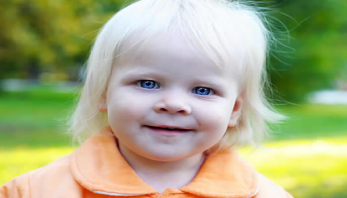
The disease is usually first suspected either because of coexistent hypopigmentation (infants born with CHS have non pigmented skin—similar to albinos but in a patchy distribution—blonde hair, and blue eyes) and a history of recurrent pyogenic infections, or on the basis of a sibling in whom the diagnosis has been previously made, or after incidental observation of giant peroxidase-positive intracellular granules on a peripheral blood smear or bone marrow examination.
Signs and symptoms that usually appear soon after birth include adenopathy, aphthae, gingivitis, hyperhidrosis, malaria, jaundice, severe and extensive pyoderma, recurrent sinopulmonary infections and fever usually unrelated to recognizable infections.
- Skin Involvement
Most patients with CHS exhibit oculocutaneous albinism Opens in new window in at least one of three sites: the skin, the hair, or the eyes. Hairs are sparse and color varies from blonde to dark brown, bur always has a silvery tint that is particularly noticeable in strong light.
Patients with CHS also have less skin pigmentation than their siblings and are susceptible to sunburns. This lack of pigmentation is also noticeable in the areolas and genitals. The pigmentary disturbance is not due to absence of melanin, but to its abnormal aggregation into giant melanosomes.
- Eye Involvement
The albinism Opens in new window in CHS is often more evident in the eyes than in the skin. Ocular alterations include a lack of pigmentation in the iris and the retina, pigmentary degeneration of the peripheral retina with progressive visual loss.
Over all, in CHS the skin is fair, the retina are pale, and the irises are translucent. Ocular involvement can also be manifested clinically by photophobia, rotator nystagmus, and an increased red reflex.
Abnormal giant melanosomes have been found in the optic cup and neural crest derived melanocytes; therefore the ocular hypopigmentation in patients with CHS is related to an ultrastructural melanosomal defect.
- Recurrent Infections
Recurrent infections affect manily the skin, respiratory tract and mucous memberanes. The most commonly involved organisms are Staphylococcus aureus, betahaemolytic streptococci, Streptococcus pneumonia, and other bacteria, fungi, and viruses.
Recurrent skin infections range from superficial pyoderma to deep subcutaneous abscess and ulcers that heal slowly and result in atrophic scars. Deep ulcerations resembling pyoderma gangrenosum have been also described.
This increased susceptibility has been attributed to the various immunologic defects observed in CHS including cellular immune deficiency, absent natural killer (NK) cytotoxic activity, altered neutrophils and monocytes numbers, diminished chemotactic responses, and delayed degranulation and intracellular killing of microorganisms.
- Neurological Abnormalities
In many persons with CHS, neurological changes appear in the lymphoproliferative phase. Progressive neurological deterioration is common in patients who survive early childhood. The adult form of CHS manifests during late childhood to young adult hood and is marked by various neurological sequelae.
The various neurological abnormalies of CHS have been mainly described in young adult patients. Among the most common are:
- signs of spinocerebeller degeneration such as clumsiness and abnormal gait, progressive parkinsonian syndrome (e.g., bradykinesia, resting tremor and oculogyric crises),
- dystonia, and peripheral neuropathy which is manifested by dysesthesias and paresthesias,
- transitory pareses (including cranial nerve palsies), and
- sensory deficit of the glove-stocking type.
The sensorineural neuropathy may begin in early childhood and progress to complete loss of muscle stretch reflexes, weakness, atrophy, and sensory deficits.
Ataxia, with broad-based gait and dysdiadochokinesia, seizures Opens in new window, and behavioral abnormalities also occur. Headaches Opens in new window or emotional lability may occur occasionally in a setting of cognitive deterioration.
The patient may become bedridden and totally incapacitated. Mental retardation Opens in new window or progressive intellectual decline have been also associated with CHS and appear to be independent of other neurological signs. The intellectual limitation may progress even after cure of the hematological manifestations with bone marrow transplantation.
- Coagulation Defects
There is usually a mild bleeding diathesis associated with CHS. Coagulation studies usually show a prolonged bleeding time, a normal platelet count, and impaired platelet aggregation with epinephrine and collagen.
This results form a platelet storage pool dense granule deficiency. Patients usually experience increased cutaneous bruising during the chronic phase, even though thrombocytopenia and severe hemorrhage can occur during the accelerated phase.
Pathogenesis and Molecular Genetics
Chediak-Higashi syndrome is inherited as autosomal recessive Opens in new window and is caused by mutation in he lysosomal trafficking regulator gene (LYST), currently named CHS1 gene, located on chromosome 1q42.1–42.2.
The defective gene encodes a clue protein to regulate the secretory processes of intracellular lysosomal vesicles and melanosomes. The CHS protein is expressed in the cytoplasm of cells of a variety of tissues and may represent an abnormality of organelles protein trafficking.
The CHS gene affects the synthesis and/or maintenance of storage/secretory granules in various types of cells: lysosomes of leukocytes and fibroblasts, dense bodies of platelets, azurophilic granules of neurtrophils, and melanosomes and melanocytes which are generally larger in size and irregular in morphology, indicating that a common pathway in the synthesis of organelles responsible for storage is affected in patients with CHS.
Diagnosis
CHS can be diagnosed based on the presence of pathognomonic anomalous giant cytoplasmic granules in neutrophils or in leukocyte precursor cells (bone marrow smears) and multiple small clumps of melanin along the hair shaft distributed in a regular pattern.
Fluorescent cytometric analysis reveals the characteristic leukocyte dysfunction, the cellular granularity and surface molecules.
Light and electron microscopy examinations of biopsy specimens of periodontal tissues reveal massive bacterial invasion of epithelial tissue, epithelial cells and connective tissue.
Prenatal diagnosis can be made by examnation of the hair from foetal scalp biopsy and from leukocytes from foetal blood samples.
Differential diagnosis include albinism, bacterial mouth infections, cutaneous T-cell lymphoma, Griscelli syndrome and pyoderma gangrenosum.
Prognosis
The prognosis of CHS is generally poor; early death occurs without treatment from infections, hemorrhage, or complications of the accelerated phase. However, there is significant clinical heterogeneity among patients with CHS, and some patients may survive into adulthood with few or even no severe infections, although they may develop progressive neurological deterioration.
Therapeutics
Current treatment protocols for CHS include prophylactic trimethoprim-sulphamethoxazole and aggressive parenteral antibiotic treatment of infections. Ascorbic acid (vitamin C) at doses of 20 mg/Kg or 0.2–6 grams per day has been shown to improve neutrophils function in vitro, although there is no proof that this provides clinical benefit.
High dose of methylprednisolone and splenectomy in the accelerated phase may improve significantly the clinical, radiological and hematological findings.
Allogeneic bone marrow transplantation from an HLA-matched sibling is the therapy of choice and should be performed early. In absence of a family donor, an unrelated donor or a placental blood graft is a good alternative.
Cure of the accelerated phase can be achieved even when transplantation results only in mixed chimerism, suggesting that even a small fraction of donor cells are sufficient to suppress this phase. Conversely, ocular and skin pigmentary disturbances are not corrected by the bone marrow transplantation and neurological complications can still develop or relapse after the transplant.
- Apitz-Castro R, Cruz MR, Ledezma E, Merino F, Ramirez-Duque P, Dangelmeier C, Holmsen H (1985) The storage pool deficiency in platelets from humans with the Chediak-Higashi syndrome: study of six patienst. Br J Haematol 59:471-483.
- Aslan Y, Erduran E, Gedik Y, Mocan H, Yildiran A (1996) The role of high dose methylprednisolone and splenectomy in the accelerated phase of Chediak-Higashi syndrome. Acta Haematol 96:105-107.
- Barrat FJ, Auloge L, Pastural E, Lagelous RD, Vilmer E, Cant AJ, Weissenbach J, Le Paslier D, Fischer A, de Saint Basile G (1996) Genetic and physical mapping of the Chediak-Higashi syndrome on chromosome 1q42–43. Am J Hum Genet 59:625-632.
- Buchanan GR, Handin RI (1976) Platelet function in the Chediak-Higashi syndrome. Blood 47:941-948.
- De Freitas GR, de Oliveira CP, Reis RS, Sarmento MR, Gasper NK, Fialho M, Praxedes H (1999) Seizures in Chediak-Higashi syndrome. Arq Neuropsiquiatr 57: 495-497.
- Farah RA, Rogers R (2004) Chediak-Higashi syndrome: In: Roach ES, Allen VS (eds.) Neurocutaneous Disorders. New York: Cambridge University Press, pp. 296-300.
- Haddad E, Le Diest F, Blanche S, Benkerrou M, Rohrlich P, Vilmer E, Griscelli C, Fischer A (1995) Treatment of Chediak-Higashi syndrome by allogeneic bone marrow transplantation: report of 10 cases. Blood 85:3328-3333.
- Hauser RA, Friedlander J, Baker MJ, Thomas J, Zuckerman KS (2000) Adult Chediak-Higashi parkisonian syndrome with dystonia. Mov Disord 15:705-708.
- Higashi O (1954) Congenital gigantism of peroxidase granules: the first case ever reported of qualitative abnormality of peroxides. Tokai J Exp Clin Med 59: 315-332.
