
Tinea Versicolor (Pityriasis Versicolor)
Definition and Cutaneous Features
|
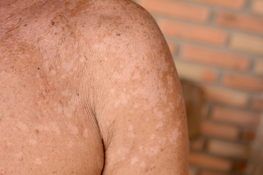
Tinea versicolor (also called pityriasis versicolor) is a superficial skin infection characterized by hypopigmented, hyperpigmented, or erythematous macular eruption which results in macules (spots darker or lighter than normal skin tone) appearing on affected area. |
Macules may coalesce into large patches with an adherent fine scale.
Macules (or spots) can appear anywhere on the body because tinea versicolor is not limited to one or two areas of the body. However, the predominant areas for pityriasis versicolor—as it's also known—is the chest, upper arms, back, or neck.
In some individuals, the lesions can be tan, salmon, brown, red, or pink. This eruption begins during adolescence, when the sebaceous glands become active.
The eruption tends to flare when the temperatures and humidity are high. Immunosuppression, systemic corticosteroids, and sweaty or greasy skin will also cause this disease to flare.
Pathogenesis
Tinea versicolor is caused by the lipophilic yeast of the genus Malassezia. This yeast is part of the normal skin flora in seborrheic areas.
The infection results from a change to the mycelia state of dimorphic lipophilic yeasts of the genus Malassezia, which colonize the stratum corneum, under the influence of various factors including moisture, heat, the degree of host immunity and inherited predisposition.
The organism produces the dicarboxylic acid, azelaic acid, which is a tyrosinase inhibitor and may contribute to the hypopigmentation noted in this infection.
The genus Malassezia was previously called Pityrosporum with two species: Pityrosporum ovale and Pityrosporum orbiculare.
Malassezia genus was initially separated into two species: the lipophilic yeast Malassezia furfur and the non-lipophilic Malassezia pachydermatis.
New species of lipophilic Malassezia besides M. furfur have been delineated: M. sympodialis, M. globosa, M. slooffae, M. restricta, and M. obtucs.
Recently on the basis of DNA relatedness, four new species, namely, M. dermatis, M. nana, M. Japanica, and M. yamatoiensis, have been recognized. Malassezia globosa, M sympodialis, and M. furfur seem to be the most common species producing tinea versicolor.
The condition is most commonly seen in adolescents and young adults but it may affect infants and even newborns.
Clinical Manifestation
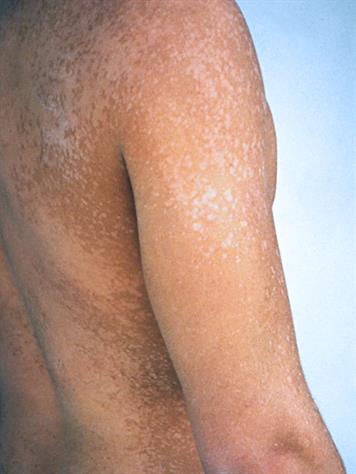
Tinea versicolor manifests with well-defined, round to oval asymptomatic, or slightly pruritic, minimally scaly hypopigmented macules that may coalesce into larger patches.
In some individuals, the lesion may be red-brown or brown. A variant with red macules (pityriasis versicolor rubra) and another with black ones (pityriasis versicolor nigra) has been described.
The lesions are more prominent in the summer months, not only because of the warm weather but also because normal skin tans while the affected areas often do not.
Noted also is that the lesions are distributed over the upper trunk, neck, and proximal upper extremities; facial lesions are not uncommon on the forehead.
Diagnosis
The diagnosis of tinea versicolor is readily made by examination of a KOH preparation of scales Opens in new window from the hypopigmented lesions that show numerous short hyphase and yeast forms.
The main differential diagnostic possibilities in tinea versicolor are post-inflammatory hypopigmentation Opens in new window and pityriasis alba Opens in new window.
Therapeutics
Treatment may be accomplished with one of the antiseborrheic shampoos such as selenium sulfide, or ketoconazole, or topical imidazole antifungal agents.
Oral ketoconazole, itraconazole or fluconazole in older children may also be effective, especially in widespread cases.
Hypopigmentation without any scale may remain for several months after treatment, even through the pathogenic organism has been eradicated. Sun exposure often evens out the pigmentation changes somewhat more quickly after treatment has been completed. Regardless of what therapy is used, recurrences are common.
See also:
- Schwartz RA. Superficial fungal infections. Lancet. 2004;364:1173-1182.
- Jung EG, Bohnert E. Mechanism of depigmentation in pityriasis versicolor alba. Arch Dermatol Res. 1976;256:333-334.
- Erchiga VC, Florencio VD: Malasseiza species in skin diseases, Curr Opin Infect Dis 15: 133-142, 2002.
- Crespo-Erchiga V, Florencio VD. Malassezia yeast and pityriasis versicolor. Curr Opin Infect Dis. 2006;19:139-147.
- Gaitanis G, Velegraki A, Alexopoulos EC, et al. Distribution of Malassezia species in pityriasis versicolor and seborrhoeic dermatitis in Greece. Typing of the major pityriasis versicolor isolate M. globosa. Br J Dermatol. 2006;154:854-859.
- Nanda A, Kaur S, Bhakoo ON, et al. Pityriasis (tinea) versicolor in infancy. Pediatr Dermatol. 1988;5:260-262.
- Maeda M, Makimura KC, Yamaguchi H, Pityriasis versicolor rubra. Eur 1 Dermatol 2002;12:160-164.
- Partap R, Kaur I, Chakrabani A, et al. Single-dose fluconazole versus itraconazole in pityriasis versicolor. Dermatology. 2004;208:55-59.
