
Nevus Anemicus
Definition and Cutaneous Features
|
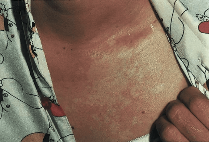
Nevus anemicus is an uncommon congenital vascular disorder in which there is usually a solitary asymptomatic patch that is paler than the surrounding normal skin. Its margin is irregular and there may be islands of sparing within the lesion. The pale area averages 5–10 cm in diameter. There is a female predominance. |
Although it occurs most commonly on the upper trunk, nevus anemicus has also been reported on the extremities and the head and neck. A variant with multiple lesions on the arms has been reported. It persists unchanged throughout life.
Extracutaneous Findings
Nevus anemicus may be seen in close association with port wine stains. These twin anomalies may be a result of somatic recombination of allelic mutations for vasoconstriction and vasodilatation.
Phakomatosis pigmentovascularis Opens in new window, where vascular and pigmented nevi occur in association with nevus anemicus, has been used to support this concept.
Lesions of nevus anemicus occur with increased frequency in patients with neurofibromatosis Opens in new window. Onset in one patient was at age 21 years, suggesting the likelihood of an acquired variant of nevus anemicus.
Biers spots Opens in new window have a similar appearance, but they are permanent, often associated with venous stasis. They probably result from anatomical or functional damage to small blood vessels.
Nevus anemicus is regarded as a pharmacological nevus in which the pallor is attributable to increased sensitivity of the blood vessels in the area to catecholamines.
It has been found that the vessels do not respond normally to proinflammatory cytokines, at least at the level of E-selectin expression. Nevus oligemicus Opens in new window is a related entity in which there is livid erythema rather than pallor.
Pathogenesis
Examination by light and electron microscopy reveals no abnormality, and the nevus is best characterized as a pharmacological abnormality, rather than an anatomical one. The pallor is due to an increased local vascular reactivity to catecholamines Opens in new window.
Donor dominance was demonstrated by grafting lesional skin from nevus anemicus to normal skin, which retained its pale appearance, emphasizing that nevus anemicus is due to increased sensitivity of the blood vessels to catecholamines rather than to increased sympathetic stimulation.
Differential Diagnosis
Under diascopic pressure with a glass microscope slide, the lesion becomes indistinguishable from the blanched surrounding skin.
Wood’s lamp Opens in new window examination does not accentuate the lesion, and rubbing or temperature change causes eryethema in the surrounding area but not within the lesion itself.
These maneuvers help to distinguish nevus anemicus from vitiligo Opens in new window, nevus depigmentosus Opens in new window, tuberous sclerosis macules Opens in new window, tinea versicolor Opens in new window, and leprosy Opens in new window.
Therapeutic
Treatment is cosmetic, and if desired the discoloration can be concealed with camouflage make-up.
See also:
- Jimbow K, Fitzpatrick TB, Szabo G, Hori Y. Congenital circumscribed hypomelanosis: a characterization based on electron microscopic study of tuberous sclerosis, nevus depigmentosus, and piebaldism. J Invest Dermatol. 1975;64(1):50-62.
